Electrocardiography is one of the most widely used methods for evaluating heart activity in modern medicine. The test records the electrical signals produced by the heart and helps doctors detect various cardiac conditions. Accurate ecg lead placement is essential for obtaining reliable results during this procedure. When electrodes are positioned correctly on the patient’s body, the electrocardiogram can clearly display the heart’s rhythm and electrical patterns. Improper placement may lead to misleading readings and incorrect interpretation. Understanding the principles of ecg lead placement helps healthcare professionals ensure accurate diagnosis, effective monitoring, and better patient care in hospitals and clinical settings.
What Is an Electrocardiogram
An electrocardiogram is a medical test that records the electrical activity of the heart over a period of time. Small sensors called electrodes are attached to specific locations on the body to detect these electrical signals. The recorded information appears as waves on a monitor or printed graph that doctors analyze for abnormalities. Accurate ecg lead placement is crucial because each electrode captures signals from a different angle of the heart. When these signals are combined, they provide a comprehensive view of heart function. This simple yet powerful diagnostic tool helps identify irregular heart rhythms, heart damage, and other cardiovascular conditions.
Importance of Correct ECG Lead Placement
Correct ecg lead placement plays a major role in ensuring the accuracy of electrocardiogram readings. Each electrode position is carefully selected to detect electrical activity from a specific direction of the heart. If leads are placed incorrectly, the recorded signals may appear abnormal even when the heart is functioning normally. This can lead to confusion during diagnosis and may require repeating the test. Healthcare professionals receive training to recognize anatomical landmarks on the body for proper electrode positioning. By maintaining precise ecg lead placement, clinicians ensure that the test reflects the true electrical behavior of the heart.
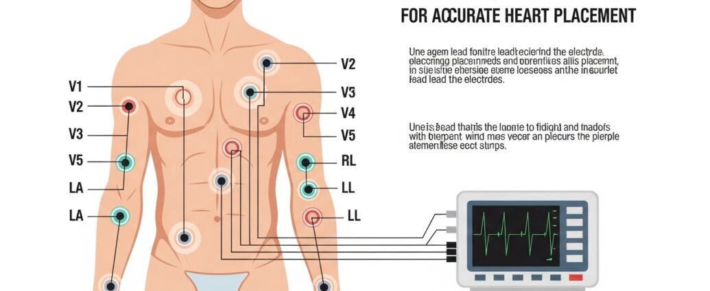
Understanding the Standard 12 Lead ECG System
The standard electrocardiogram commonly used in hospitals is the 12 lead ECG system. This method uses ten electrodes placed on the body to generate twelve different views of the heart’s electrical activity. These leads provide detailed information about various regions of the heart, including the front, sides, and lower portions. Proper ecg lead placement is essential to capture accurate signals from each of these perspectives. The combination of limb leads and chest leads allows doctors to study the heart’s rhythm, detect blockages, and identify signs of heart attack or other cardiac disorders with greater precision.
Limb Lead Placement in ECG Testing
Limb leads are an important component of ecg lead placement because they help record electrical signals from the heart through the arms and legs. Electrodes are typically attached to the right arm, left arm, right leg, and left leg. These positions form the basis for measuring electrical activity in different directions across the heart. The limb leads create several views that assist doctors in evaluating heart rhythm and conduction patterns. Correct placement ensures that electrical signals travel accurately through the monitoring system. Even slight variations in electrode position can affect the recorded waveform and influence medical interpretation.
Chest Lead Placement and Its Significance
Chest leads provide detailed information about the heart’s electrical activity from the front of the chest. These electrodes are placed at specific locations across the chest wall to capture signals from different areas of the heart muscle. Accurate ecg lead placement for chest leads allows doctors to observe how electrical impulses travel through the ventricles. This information is essential for identifying conditions such as heart attacks, ventricular enlargement, and conduction abnormalities. Proper spacing and positioning of chest electrodes ensure that each lead records a clear and distinct signal, which improves the overall reliability of the electrocardiogram.
Preparing the Patient for ECG Lead Placement
Proper patient preparation is a key step before performing an electrocardiogram. Healthcare professionals ensure that the patient is comfortable and relaxed while lying in a stable position. The skin where electrodes will be attached is usually cleaned to remove oils or sweat that may interfere with signal detection. Sometimes small areas of hair are removed to improve electrode contact. These steps help maintain accurate ecg lead placement and allow the electrodes to transmit electrical signals effectively. Careful preparation also reduces movement artifacts, which can distort ECG recordings and make it harder for doctors to interpret the results.
Common Errors in ECG Lead Placement
Despite its routine nature, ecg lead placement errors can occur if proper attention is not given during the procedure. One common mistake involves placing chest leads in incorrect intercostal spaces or shifting limb electrodes from their standard positions. These errors may alter the appearance of ECG waveforms and mimic serious heart conditions. Misplaced electrodes can create patterns that resemble heart attacks or conduction blocks. Healthcare professionals must remain attentive to anatomical landmarks to avoid such issues. Recognizing and correcting placement errors helps maintain the accuracy of ECG results and prevents unnecessary medical concern.
Role of ECG Lead Placement in Cardiac Diagnosis
Accurate ecg lead placement is essential for diagnosing a wide range of heart conditions. Doctors rely on ECG readings to identify arrhythmias, heart attacks, electrolyte imbalances, and structural abnormalities. Each lead offers a unique view of the heart’s electrical activity, allowing physicians to pinpoint the location of potential problems. When electrodes are positioned correctly, the ECG provides reliable data that guides medical decision making. This information supports early detection and timely treatment of cardiovascular diseases. Proper lead placement therefore plays a direct role in improving patient outcomes and supporting effective cardiac care.
Training and Skills Required for Accurate Placement
Healthcare professionals who perform electrocardiograms receive specialized training to master ecg lead placement techniques. Nurses, technicians, and medical assistants learn how to identify anatomical landmarks and apply electrodes correctly for both limb and chest leads. Training also includes understanding how body shape, posture, and patient movement may affect electrode positioning. Experience helps clinicians perform the procedure quickly while maintaining accuracy. Continuous education ensures that healthcare workers remain familiar with best practices in electrocardiography. Skilled placement not only improves the quality of ECG recordings but also enhances confidence in the diagnostic information obtained from the test.
Conclusion
Accurate ecg lead placement is fundamental to obtaining reliable electrocardiogram results and ensuring proper evaluation of heart function. Each electrode position provides valuable information about the heart’s electrical activity from different angles. When healthcare professionals follow correct placement techniques and prepare the patient properly, the ECG becomes a powerful tool for diagnosing cardiac conditions. Errors in placement can lead to misleading readings, making precision essential during the procedure. By understanding the importance of electrode positioning and maintaining careful attention to detail, medical professionals can use electrocardiography effectively to support accurate diagnosis and improved patient care.